FIG 7.

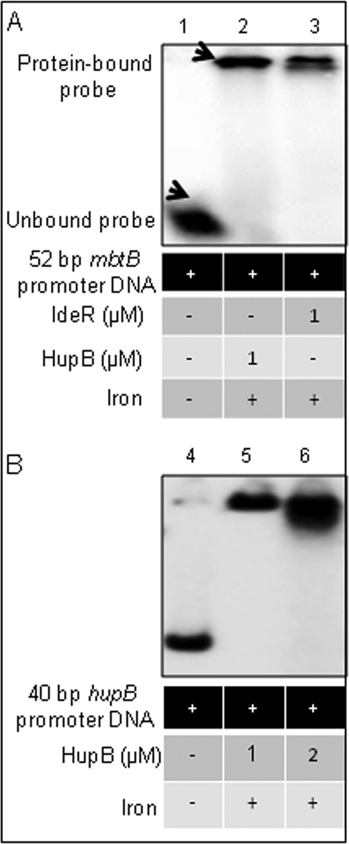
EMSA confirmation of the binding of HupB to the 10-bp AT-rich HupB box. (A) A chemically synthesized 52-bp mbtB promoter region, containing the IdeR box and the HupB box, was labeled with [γ-32P]ATP and subjected to EMSA (as done earlier; iron was added at 200 μM) in the presence of HupB (lane 2) and IdeR (lane 3); lane 1 served as a control showing the unbound probe in the absence of any added protein. (B) To confirm further the binding of HupB to the 10-bp HupB-binding motif, a 40-bp oligonucleotide containing the putative HupB box in the hupB promoter (identified by in silico analysis; Table 4) was subjected to EMSA. Lane 4 shows the unbound probe, and lanes 5 and 6 show increasing amounts of the bound 40-bp probe upon addition of increasing amounts of the HupB protein.
