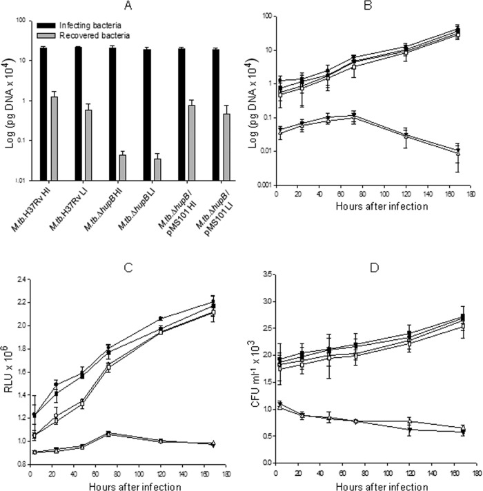
FIG 8.
Infectivity and survival of M. tuberculosis H37Rv, M. tuberculosis ΔhupB mutant, and the hupB-complemented M. tuberculosis ΔhupB/pMS101 strains inside macrophages. Cells (106) of the mouse peritoneal macrophages (RAW 264.7 cell line) seeded in a 6-well plate were infected with high-iron-grown M. tuberculosis H37Rv (●), M. tuberculosis ΔhupB mutant (▼), and M. tuberculosis ΔhupB/pMS101 (■) strains and the respective low-iron-grown organisms (M. tuberculosis H37Rv, ○; M. tuberculosis ΔhupB mutant, Δ; M. tuberculosis ΔhupB/pMS101, □) at an MOI of 10:1. (A) The number of cells recovered from the macrophages 4 h after infection (determined by qRT-PCR) was considered the number of cells that gained entry into the macrophages (and time zero for studying the viability inside macrophages), and subsequent isolation of bacilli from the macrophages was done on days 1, 2, 3, 5, and 7. The number of viable bacilli in each of the wells was assayed by both ATP assay (B) and qRT-PCR analysis based on 16S rRNA (C); this was assayed in triplicate. Two such identical experiments were done for each of the M. tuberculosis ΔhupB mutant and M. tuberculosis ΔhupB/pMS101 strains. (D) The number of viable bacilli determined as CFU.

