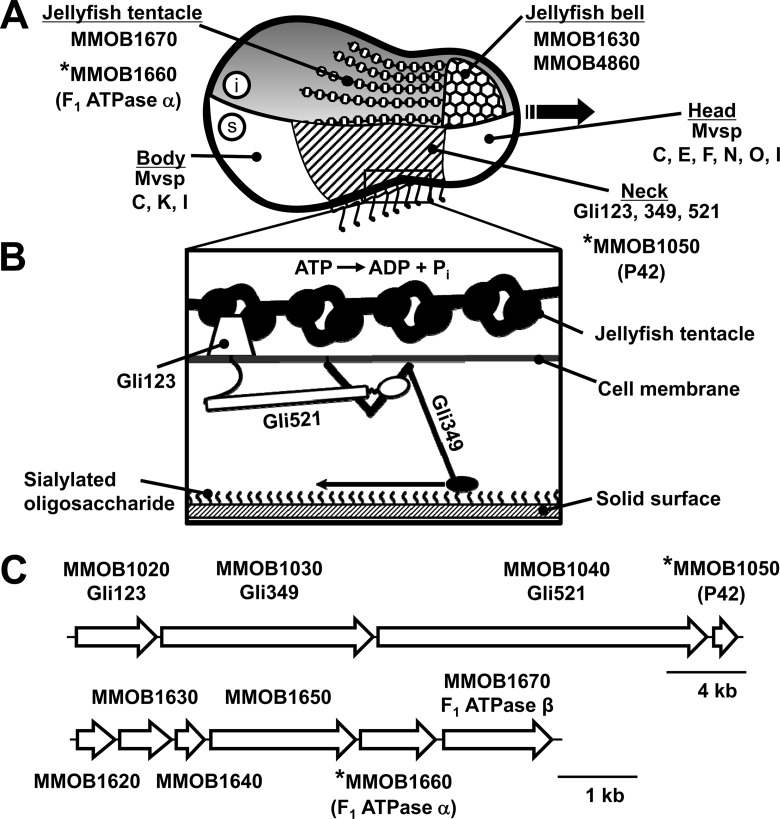
FIG 1.
Schematic of M. mobile cell architecture (2, 3, 55). The ORFs considered in this study are marked by asterisks. (A) Drawing of the cell showing the inside structure (i, above) and the surface structure (s, below). The cytoskeletal jellyfish structure can be divided into two parts, the “bell” and the “tentacles.” The bell is composed of protein products of the MMOB1630 and MMOB4860 genes, and the tentacles are protein products of the MMOB1670 gene. The cell surface can be divided into three parts, the head, neck, and body, beginning at the front end. The gliding direction is indicated by the black arrow. The neck is covered by the Gli123, Gli349, and Gli521 proteins, all of which are essential for gliding. The head is covered with MvspC, -E, -F, -N, -O, and -I, and the body is covered with MvspC, -K, and -I; these proteins are likely involved in a mechanism for evading the host immune system. (B) Magnified image of the neck surface based on our previous studies, most of which employed electron microscopy. The distal globular part of Gli349 is thought to catch and pull sialylated oligosaccharides fixed on the host surface, a mechanism that hydrolyzes ATP. (C) ORFs for surface and jellyfish structures (above and below, respectively). Ten ORFs are known to encode components of the jellyfish structure, but four are located at other regions of the genome.

