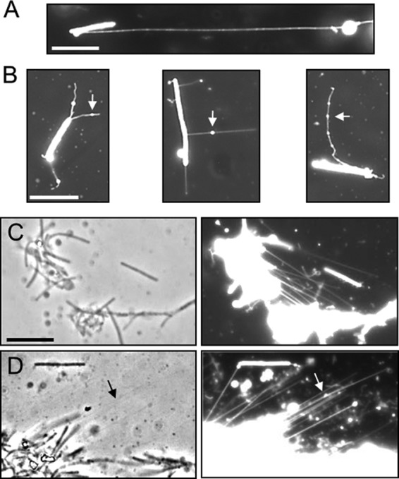
FIG 2.

OMTs detected by lipophilic dye (DilC) fluorescent staining. (A) An OMT that exceeds the length of the cell >6-fold (DK8601; aglB1 ΔpilA). (B) Vesicle-like structures (arrows) associated with OMTs (DW1047). (C) Two clumps of cells appear to be connected by OMTs (compare left and right panels) (DK8615; ΔpilQ). (D) OMT bundles are faintly visible by phase contrast (arrow) and appear to anchor a cell clump to the glass slide (right panel; DK8615). Bars, 8 μm.
