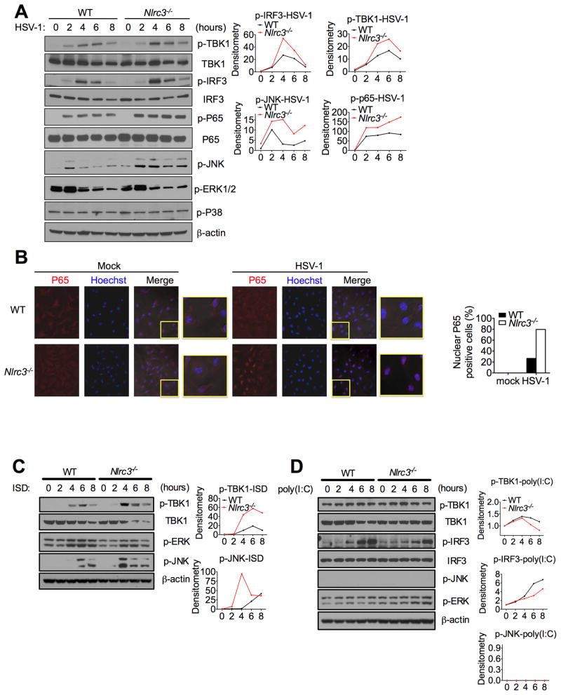
Figure 6. NLRC3 deficiency enhances immune signaling.
(A) Immunoblot of phosphorylated (p-) TBK1, IRF3, p65, JNK, ERK and p38 in lysates of WT and Nlrc3−/− MEFs infected with HSV-1 (MOI 1) for indicated time points. Densitometric measurements are depicted to the right. (B) BMDMs isolated from WT or Nlrc3−/− mice were infected with HSV-1 (MOI 1) for 2.5 hours. Cells were fixed and stained for endogenous p65 (red) or hoechst which stains the nucleus (blue). The merged purple color is indicative of nuclear p65. (C) Similar to (A), except cells were transfected with ISD. (D) Similar to (A) except cells were transfected with poly(I:C). Data are representative of at least two independent experiments.

