
Figure 7. Loss of IRS-1/2 expression results in reduced bud size and disrupted mesenchyme at E14.5.
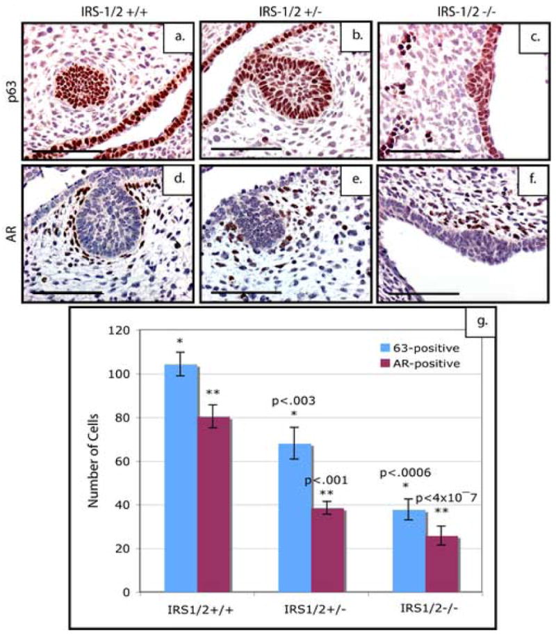
p63 expression in the epithelium of E14.5 mammary buds of (a)IRS-1/2+/+, (b) IRS-1/2+/−, (c) IRS-1/2−/− mice. Note the dramatic reduction in the number of epithelial cells that stain for p63 both in the heterozygous (b) and IRS-1/2-deficient mammary placodes (c). Note the dramatic reduction of AR-positive cells within the condensed mesenchyme surrounding the epithelial buds in (e) heterozygous and (f) deficient anlagen as compared to the wildtype. Quantitation of these staining patterns (g) show a statistically significant decrease in the number of p63-positive cells with in bud of the IRS-1/2 heterozygous and deficient embryos as well as a statistically significant decrease in the number of AR-positive cells within the p190-B heterozygous and deficient anlagen as compared to the wildtype. (f). Scale bar 100 μm.