
Fig. 2.
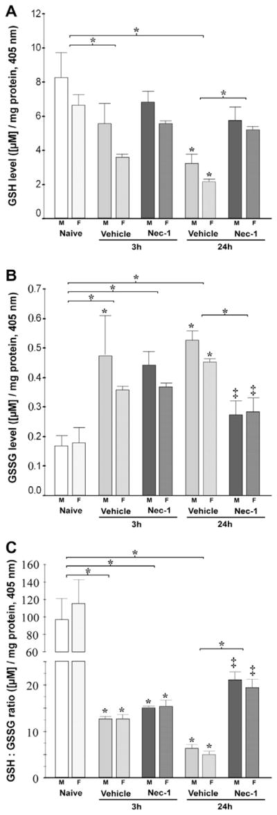
Nec-1 treatment following neonatal HI prevents early glutathione oxidation in the forebrain of male and female mice. Bar graphs showing (A) reduced glutathione (GSH) levels, (B) oxidized glutathione (GSSG) levels, and (C) GSH:GSSG ratio in ipsilateral forebrain at 3 and 24 h following neonatal HI. Bars represent the mean±SEM measured in naive control (white), vehicle (light gray) and Nec-1 (dark gray) treated male (solid color, M) and female (hashed color, F) mice. p ≤ 0.05 in all cases (one-way ANOVA). *, vs. naive control; ‡, vs. 24 h vehicle; p<0.05 (Tukey’s post hoc). n=4–5 mice/treatment/ time/gender. Bracket and *, represent p ≤ 0.05 for the whole pool.