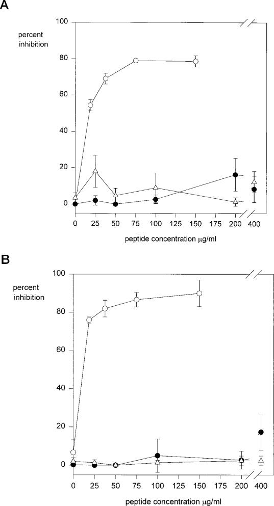
Fig. 3. Peptide inhibition of cell adhesion to region II-plus peptides.
MG63 cells (panel A) or K562 cells (panel B) were preincubated with inhibitor peptides PCSVTCGNGIQVRIKPGSAN (open circles); CSVTCG, cysteines blocked (open triangles); CSVTCG, cysteines reduced (closed circles); for 30 min at 37 °C and then plated in wells coated with 1.25 μg/ml region II-plus peptide (PCSVTCGNGIQVRIKPGSAN) for 1 h. Bound cells were quantified with crystal violet. Each point was performed in triplicate, and shown is the percent inhibition of binding compared with controls with no inhibitor.

