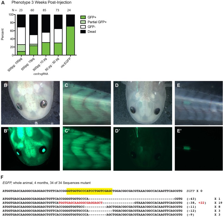
Fig. 1.
Efficient targeted mutagenesis of EGFP in Tg(CAGG:EGFP) transgenic axolotls. (A) Distribution of phenotypes in axolotl (Ambystoma mexicanum) embryos from a mating between a wild-type and hemizygous Tg(CAGG:EGFP) animal injected with various concentrations of cas9 mRNA and an EGFP-directed sgRNA at 3 weeks post-injection, as assessed by fluorescence microscopy. Whereas ∼50% of embryos injected with lower concentrations of cas9 and sgRNA displayed normal EGFP expression, those injected with the highest concentrations of cas9 and sgRNA displayed mosaic EGFP expression. Any animal exhibiting clones of EGFP-negative cells was classified as ‘partial EGFP’. The survival rate in all cas9 and sgRNA-injected embryos did not differ from that of embryos injected with nls-EGFP mRNA only (right column). (B-E′) Whereas 6-month-old uninjected Tg(CAGG:EGFP) animals display strong uniform EGFP expression in both their heads (B′) and tails (C′), siblings injected with EGFP-directed sgRNA and cas9 (D,E) display a dramatic loss of EGFP, with one individual with only EGFP-positive cells apparent in the head (D′) and another with EGFP expression only in the tail (E′). (B-E) Brightfield; (B′-E′) EGFP fluorescence. (F) All 34 sequences of cloned PCR products of the EGFP locus in a single animal injected with an EGFP-directed sgRNA and cas9 that displayed no apparent EGFP expression contain indels at the targeted site (yellow). The size of each deletion (–) or insertion (+; in red) and frequency of occurrence among clones are indicated.

