
Fig. 2.
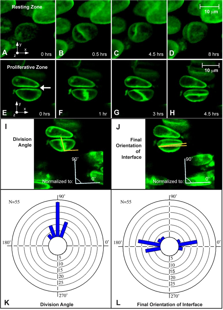
Time-lapse imaging uncovers rotation of the daughter cell interface during column formation. Time-lapse movies of three-dimensional reconstructions of dividing chondrocytes show differential behavior in the resting and proliferative zones. (A-D) In the resting zone, mitosis is oriented perpendicular to the long axis of the cell (B), and the rotation of the mitotic plane (when any occurs) is slight and disoriented (C,D). (E-H) In the proliferative zone, time-lapse movies confirm the previously demonstrated mitotic division bias parallel to the long axis of the growth plate (F). The daughter cells then remain closely associated with one another while their junction rotates to stack the cells into the expanding clonal column (G,H). The arrow in E indicates the gap between two cells in the same column created by the buildup of the pericellular matrix prior to imaging. (I,J) The angles of division and the final orientation of the interface between daughter cells were quantified by defining the x-axis as perpendicular to nearby, already-formed columns. (K,L) A strong division angle bias towards 90° was observed, and the final orientation demonstrated a bimodal distribution biased towards either 0° or 180° shown by circular histograms.