Fig. 6.
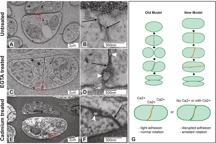
Treatment with EGTA or cadmium chloride physically disrupts daughter cell adhesion. (A-F) Transmission electron micrographs of recently divided untreated (A,B), EGTA-treated (C,D) and cadmium chloride-treated (E,F) chondrocytes show that treatments increase the distance between neighboring cell membranes at the edges of the interface (arrows; compare B with D and F). Arrowheads indicate cellular protrusions between neighboring cells not found in untreated chondrocytes. Red boxes in A, C and E indicate the area shown at higher magnification in B, D and F, respectively. (G) Based on the data presented here, the old model of division, separation and intercalation to form columns is replaced by a transient adhesion interface (red line) that is required for column formation.

