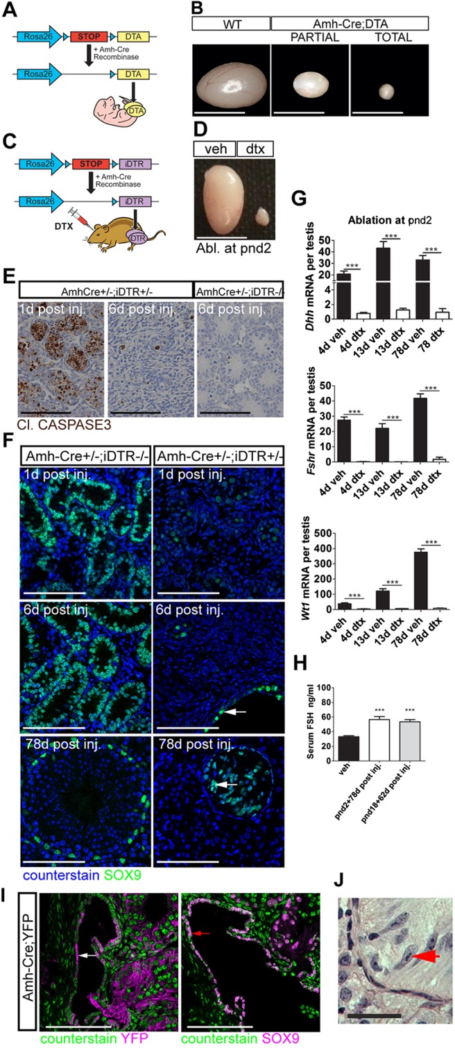
Fig. 1.

SC-specific ablation. (A) Mouse model one: Amh-Cre:DTA. (B) DTA induced testicular atrophy with different degrees of severity. (C) Mouse model two: Amh-Cre:iDTR. (D) DTX induced testicular atrophy in adulthood following injection at pnd2. (E) Apoptosis is restricted to SCs and is resolved 6 d post DTX injection. (F) SC ablation is mirrored by decreased SC SOX9 expression. Note that the expression of SOX9 is retained in the rete testis epithelium (arrows). Scale bar: 100 µm. (G) Relative expression of the SC-specific markers Dhh, Fshr and Wt1 (one-way ANOVA, n=7-9; ***P<0.001). (H) Circulating FSH concentrations at pnd80 (one-way ANOVA, n=7-9; ***P<0.001). (I) Immunolocalisation of YFP (white arrow), and SOX9 (red arrow) in Amh-Cre:YFP testis (d17). (J) Rete testis epithelium with tripartite nucleolus consistent with an SC origin for these cells (red arrow). WT, wild type; Veh, vehicle control. Scale bars: 500 µm in B,D; 100 µm in E,I; 50 µm in F,J.
