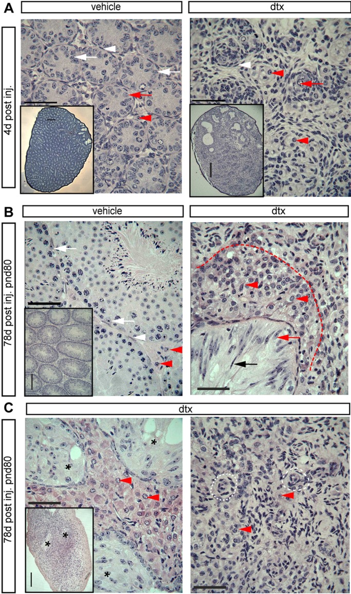
Fig. 3.

Testicular histology following SC ablation at pnd2. (A) Testicular histology of testes 4 d after ablation at pnd2. In DTX-treated animals no intact seminiferous tubules are present, although the rete testis remains present. In control testes, primordial germ cells (red arrows) and SCs (white arrows) are present inside the tubules and are surrounded by an intact PTMC layer (white arrowheads). FLCs (red arrowheads) are apparent in the interstitial tissue. In DTX-treated animals the tubules appear to have collapsed with the PTMCs forming concentric rings (white arrowheads), occasionally surrounding a surviving spermatogonial stem cell (red arrow). LCs remain present in the tissue between the collapsed tubules (red arrowheads). The insets, which are at lower magnification, show the overall structure of the seminiferous tubules in vehicle- or DTX-treated testes. (B) Testicular histology of adult testes (80 d) after ablation at pnd2. Representative SCs (white arrows), PTMCs (white arrowheads) and LCs (red arrowheads) are highlighted in vehicle-treated testis. In DTX-treated animals there was no tubular structure, although the rete testis remained intact (black arrow). The epithelium of the rete testis either had an SC-like appearance or had a highly elongated, pseudostratified appearance (black and red arrows). Abundant LCs were present in the vicinity of the rete testis (red arrowheads and delineated by the red dashed line), but there was a sharp reduction in LC numbers further from the rete testis. (C) Some variation between animals was seen in the size of the LC population surrounding the rete testis (black asterisks). In the parenchyma of the testis, concentric circles of cells were seen that might represent collapsed tubules seen at pnd6 (white dashed line). LCs were present but scarce in this region (red arrowheads). Scale bars: 50 µm in A-C; 250 µm in insets.
