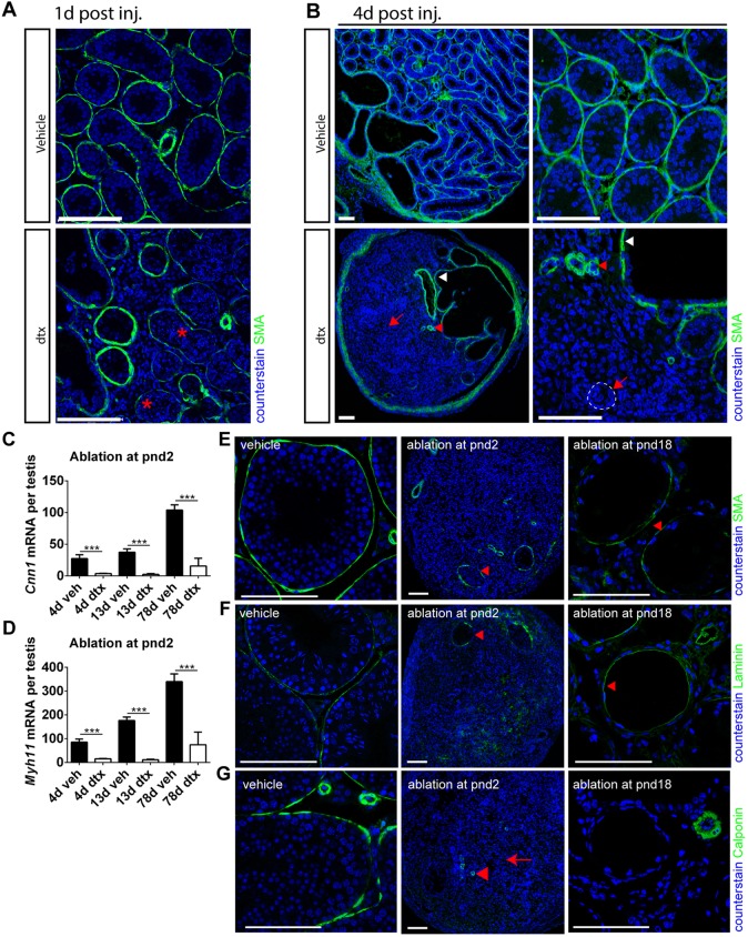
Fig. 4.
SCs maintain the PTMC differentiated phenotype in prepubertal life. (A) Disruption of PTMCs (as indicated by loss of SMA, asterisks) 1 d post DTX injection at pnd2. (B) Four days after injection, loss of SMA expression is consistent with a collapse of the tubular architecture (arrows and dashed line). Note that SMA expression is also restricted to blood vessels (red arrowhead) and rete testis (white arrowhead). (C,D) Expression of myoid cell markers (C) Cnn1 and (D) Myh11 following SC ablation at pnd2 (one-way ANOVA, n=7-9, ***P<0.001). (E) SMA expression is retained if SC ablation occurs at pnd18 (arrowhead). (F) Disruption to tubular basement membrane (BM) (laminin) at pnd2, whereas rete testis BM remains intact (arrowhead), whereas there is retention of gross tubule morphology from pnd18 (arrowhead). (G) Loss of calponin expression, a functional marker of PTMCs, at both pnd2 [arrow; whereas blood vessels retain it (arrowhead)], consistent with a collapse of the tubular architecture, and at pnd18 albeit with retention of gross tubule morphology. Scale bars: 100 µm.

