Fig. 3.
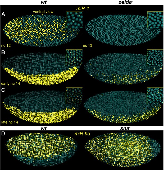
Patterning factors spatially regulate miRNA expression. Pseudocolored confocal FISH images of embryos hybridized with pri-miR-1 (A-C) or pri-miR-9a (D) probes. (A-C) Wild-type (left) and zelda mutant (right) embryos; insets show the actual FISH signal localized in nuclei. Note that the expression of miR-1 was first detected in nuclear cycle (nc) 12 in wt (A, left), but was not detected until early nc 14 in zelda mutants and the expression was also less robust (B, right). (D) miR-9a expression expands ventrally in sna mutants (right), indicating that Sna represses miR-9a in the presumptive mesoderm.

