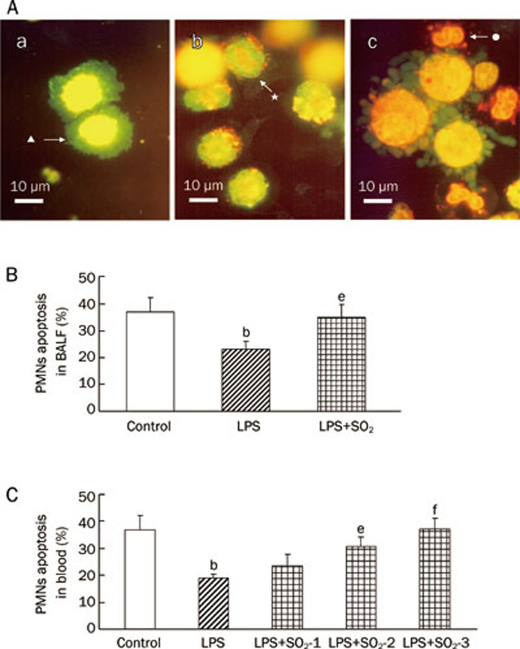
Figure 3.
PMN apoptosis determined by AO/EB-staining and FCM at 6 h after induction of ALI with or without SO2 pretreatment. (A) PMN apoptosis determined by AO/EB-staining in BALF. a) No apoptotic PMNs, only alveolar macrophages (▴), were observed in the lungs of control rats; b) Only a few early apoptotic PMNs (★) were observed (strong nuclear green fluorescence staining) in the LPS group. c) A large number of apoptotic PMNs (•) were found in the lungs of rats pretreated with SO2 before LPS administration (LPS+SO2) rat. Condensed and fragmented nuclear were stained by orange EB and apoptotic PMNs in LPS+SO2 group. Scale bar, 10 μm. (B) The percentage of apoptotic PMN cells in BALF, as determined by flow cytometry. Control, control group; LPS, LPS administration group; LPS+SO2 refers to SO2 pretreatment before LPS administration. (C) The percentage of apoptotic PMNs in the peripheral blood, as determined by flow cytometry. SO2-1, pretreatment with 10 μmol/L SO2;SO2-2, pretreatment with 20 μmol/L SO2;SO2-3, pretreatment with 30 μmol/L SO2. Because PMNs were absent in the BALF of the control group, we replaced BALF with peripheral blood for the control group. Data are presented as the mean±SD (n=7 in each group). bP<0.05 compared with the control group; eP<0.05, fP<0.01 compared with the LPS group.

