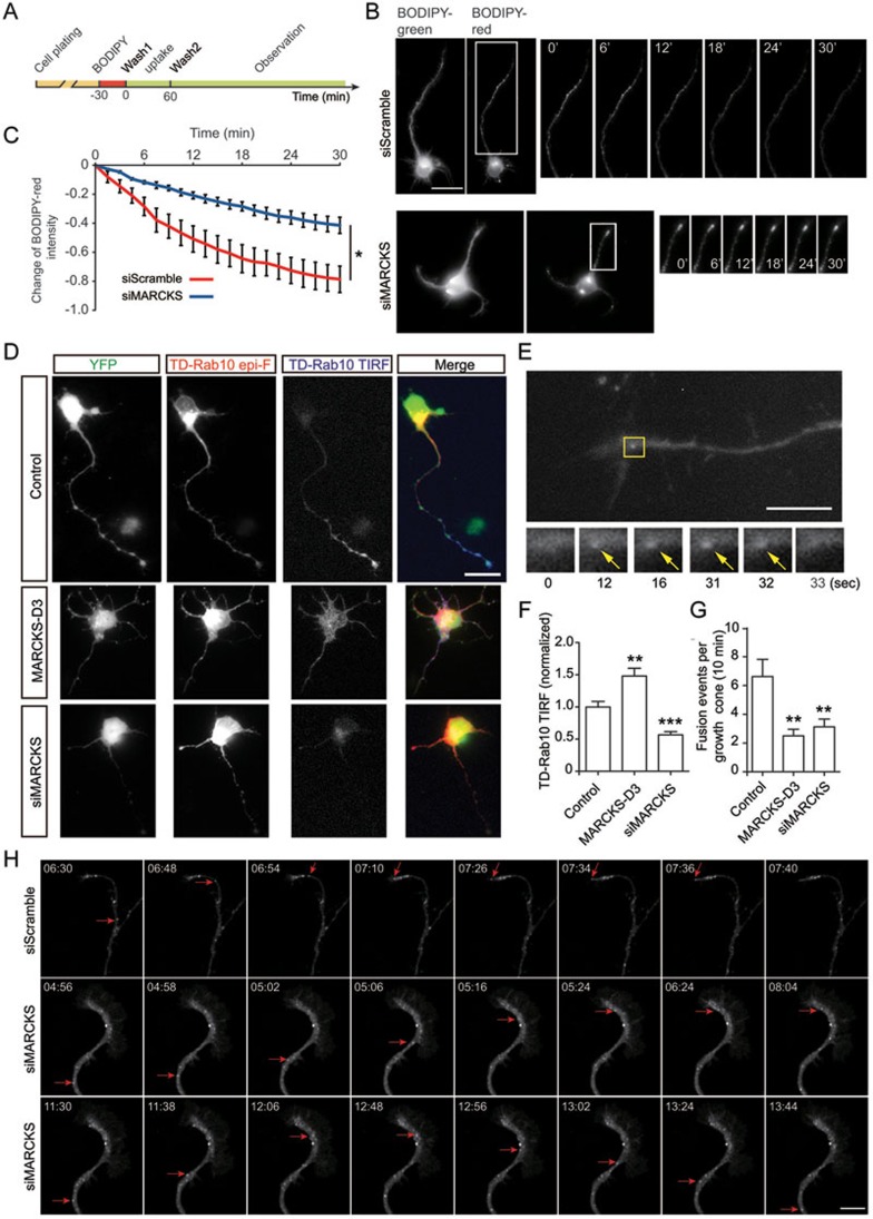
Figure 5.
MARCKS is important for membrane insertion of Rab10 vesicles in primary neurons. (A) Schematic diagram illustrating BODIPY-ceramide labeling and observation procedure. (B, C) Cortical neurons transfected with vector encoding siMARCKS or scrambled sequences were labeled with BODIPY-ceramide at DIV2. The changes of BODIPY-red signals in the longest neurite were recorded using live-imaging fluorescence microscopy. At least five cells in each group were analyzed. *P < 0.05, paired t-test. Scale bar, 20 μm. (D) Cortical neurons were co-transfected with TD-Rab10 and pEYFP-N1 (control), MARCKS-D3-YFP, or pSUPER vector encoding siMARCKS. Epifluorescence (epi-F) and TIRF signals represent total and PM-associated TD-Rab10, respectively. Scale bar, 25 μm. (E) Live imaging of Rab10 TIRF signals in axonal growth cones of DIV2 cortical neurons transfected with TD-Rab10 and various plasmids. Shown is a representative image. Arrows point to a fusion event. Scale bar, 10 μm. (F) Quantification of the TIRF signals relative to epi-F signals. Average values from control group were normalized as 1.0. Data are presented as mean ± SEM. 10 neurons in each group were analyzed. **P < 0.01, ***P < 0.001, one-way ANOVA. (G) Quantification of the frequency of vesicle fusion in axonal growth cones. Data are presented as mean ± SEM. 10 neurons in each group were analyzed. **P < 0.01, one-way ANOVA. (H) Snapshots of time-lapse images showing the movement of TD-Rab10 vesicles in axonal growth cone of DIV3 neurons transfected with siScramble or siMARCKS. Note the fading of transporting vesicles in axonal tip of control cell and persistence or retrograde transport in siMARCKS cells (red arrows). Scale bar, 10 μm. See also Supplementary information, Movies S6 and S7.

