Figure 2.
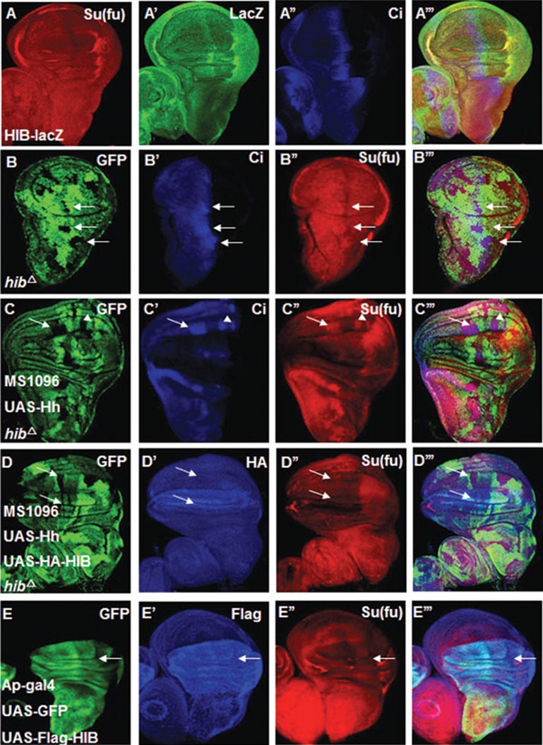
Hh signaling downregulates Su(fu) through HIB. (A-A''') LacZ expression in wing discs from a hib enhancer trap line, l(3)03477 (hib-Z). High LacZ staining (green) overlapped with low Su(fu) (red) region along the A/P border. (B-B''') Su(fu) (red) was upregulated in the hibΔ clones near the A/P boundary. hibΔ clones were marked by the lack of GFP expression and were indicated by arrows. (C-C''') A wing disc carrying hibΔ clones and expressing UAS-Hh with MS1096 was immunostained with GFP (green), CiFL (blue) and Su(fu) (red) antibodies. Su(fu) and CiFL accumulated in hibΔ clones when Hh was overexpressed. (D-D''') Co-expression of UAS-Hh and UAS-HA-HIB (blue) with MS1096 could erase the accumulated Su(fu) (red) inside hibΔ clones of wing discs. (E-E''') Wing discs expressing UAS-Flag-HIB plus UAS-GFP with Ap-Gal4 were immunostained with Flag (blue), GFP (green) and Su(fu) (red) antibodies. GFP marks the cells that express UAS-Flag-HIB. HIB overexpression led to diminishing Su(fu) level in both A- and P-compartment cells in the dorsal region of wing pouch (indicated by arrows).

