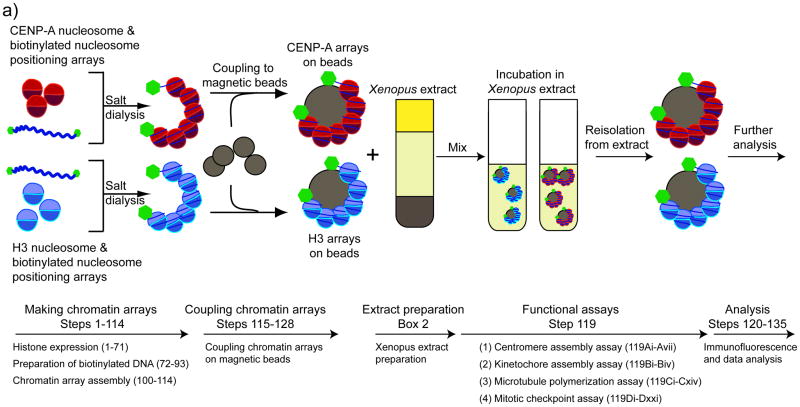
Figure 1. A cell free system for centromere and kinetochore reconstitution.
A schematic showing the general experimental procedure and the corresponding steps in the protocol. CENP-A and H3 chromatin arrays are reconstituted from purified proteins and biotinylated high affinity nucleosome positioning sequences using the salt dialysis method. Assembled chromatin arrays are coupled to Streptavidin-coated magnetic beads. The chromatin arrays are incubated in Xenopus egg extracts. After recovery, centromere and kinetochore assembly and function is analyzed. (Figure is adapted from 2).

