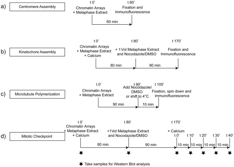
Figure 2. The cell free system can be used to analyze centromere assembly, kinetochore assembly, microtubule polymerization and mitotic checkpoint activation.
A schematic showing the four different types of assays described here analyzing the ability of reconstituted chromatin arrays coupled to beads to (A) assemble centromeres in CSF extracts (Step 119A) (B) assemble kinetochores in cycled extracts (Step 119B), (C) promote microtubule polymerization and stabilization in CSF extracts (Step 119C) and (D) activate the mitotic checkpoint in cycled egg extracts (Step 119D).

