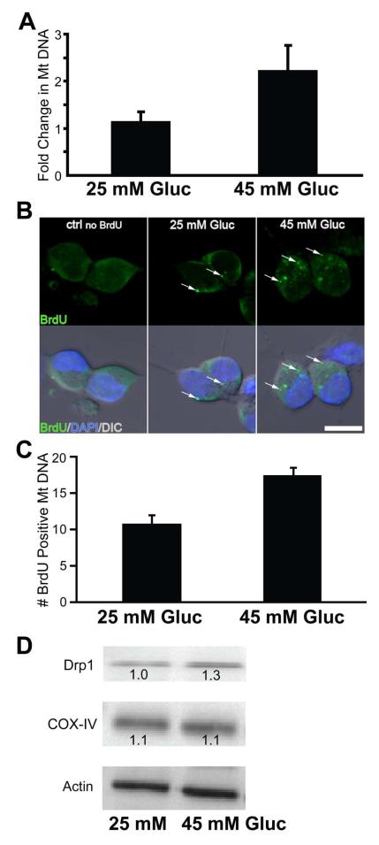
Fig. 5. Hyperglycemia increases mitochondrial biogenesis and fission in DRG neurons in vitro.
(A) DRG. Dissected and dissociated rat E15 DRGs were incubated in the presence of control (25 mM) or high glucose media (45 mM) for 6 h. Quantitation of cytochorome b DNA was used as a marker for MtDNA. n=3 (B) In vitro analysis of BrdU incorporation into MtDNA of DRG neurons under hyperglycemia. DRG neurons were cultured in the presence (or absence, ctrl no BrdU) of BrdU and normal (25 mM) or high glucose (45 mM) for 6 h. BrdU incorporation into MtDNA (arrows) was visualized by immunocytochemstry with tyramide amplified AlexaFluor 488 green signal (top panels) and merged with nuclear staining (DAPI, blue) and differential interference contrast (DIC) images (lower panels). Cells were visualized using an Olympus FluoView 500 laser scanning confocal microscope. Bar = 10 μm. (C) Quantitation of MtDNA was done by identifying BrdU-positive DRG soma. In vitro cultures were incubated in control or high glucose media for 6 h. n=10 per group (D) Western blot analysis of in vitro cultures DRG after control or treatment media exposure. Protein lysates were subjected to SDS-PAGE and analyzed with antibodies against COX-IV, Drp1, and actin (internal control).

