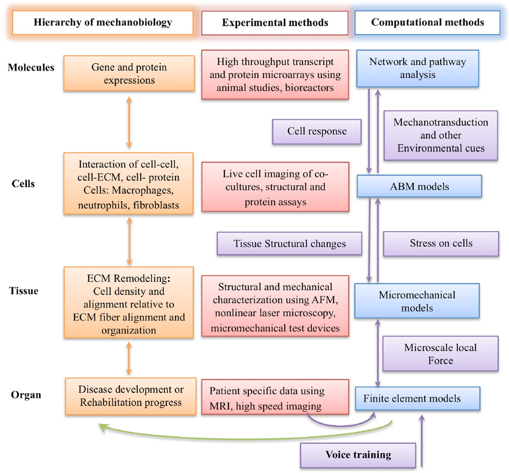
Figure 1.
Systems biology of the vocal fold mechanobiology. At the organ level, finite element modeling is used to predict the average mechanical stress and strain in vocal fold tissue. Subject specific information, such as vocal fold geometry, lamina propria structural organization and high-speed imaging can provide information for the modeling and validation of a finite element model capable of predicting average stress and strain in the tissue down to a level where continuum assumptions are no longer valid. At the tissue level, structural and mechanical characterization of tissue constituents provides inputs to micromechanical models to predict the amount of stress on a single cell. The mechanical stress on a single cell can induce an inflammatory response involving other cell types. Co-culture of cells inside ex vivo vocal fold tissue can elucidate the complex interaction between cells as well as their migration speed and dynamics, which are the essential data for the agent-based models (ABMs). In addition to the rules measureable at the cellular level using microscopic techniques, the cues that initiate the migration of these cells are the result of complex interactions inside the cells. Bioreactors can provide sufficient RNA and protein samples for genomic and proteomic analysis respectively for complex cell-cell or cell-protein interactions. The use of computer science-based pattern recognition techniques, such as pathway and network analysis, can identify and predict events at the cellular level. Integration of all the data at the molecule, cell, tissue and organ levels can constitute a multi-scale model of vocal fold mechanobiology. The clinical application of such a model is, for example, to predict the biological effect of phonation or specific voice training on vocal fold injury and healing.

