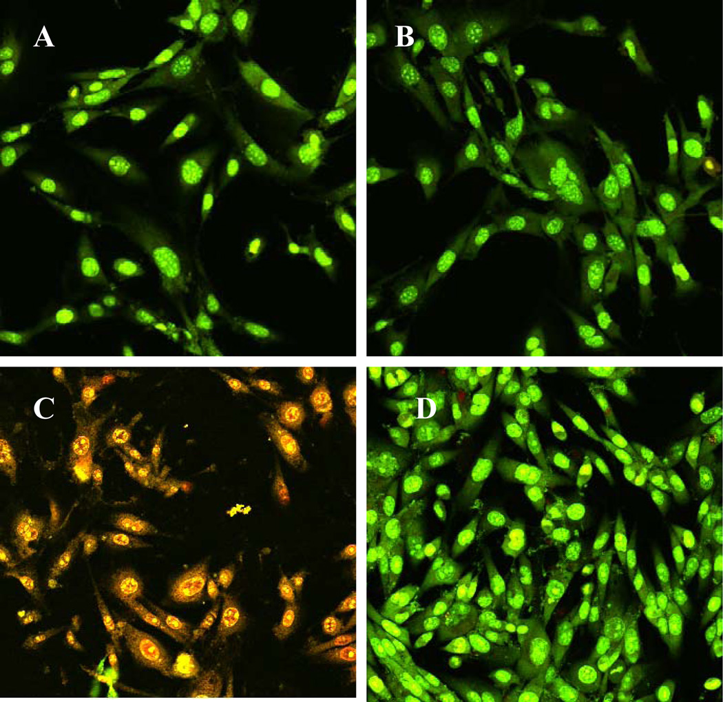
Figure 9.
Confocal microscopy images of NIH/3T3 cells on A) uncontaminated and untreated surface; B) uncontaminated and plasma treated surfaces; C) contaminated with S. aureus bacteria with no plasma treatment; and D) contaminated with S. aureus bacteria with plasma treatment (Argon discharge gas at 60 watts for 25 min and then 100 watts for 6 minutes. S. aureus suspension (2×10³/ml) was loaded onto each surface and incubated at room temperature for 2 h. At the end of the incubation time, each surface was washed in order to remove loosely adhered bacteria. Then NIH/3T3 cells in antibiotic free medium were seeded and incubated at 37 °C for an additional 6 hrs. At the end of incubation, cells were stained using Live/Dead kit.

