
FIG. 3.
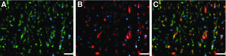
Representative images of engineered vascular grafts stained for α-smooth muscle actin (α-SMA) (green, A) and calponin (red, B). Nuclei were counterstained with DAPI (blue). Merged image is shown in (C). Scale bar, 25 μm. Color images available online at www.liebertpub.com/tea