
FIG. 4.
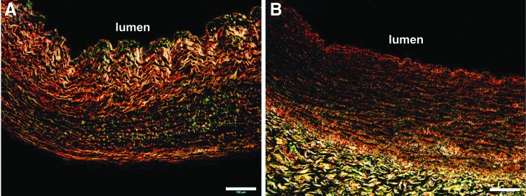
(A) Picrosirius red stain of tissue-engineered vascular graft. Red or bright orange bands (thicker collagen fiber) are toward the lumen while green bands (thinner collagen fibers) are localized at the outer region. (B) Picrosirius red stain of native bovine coronary artery shown as a control. Scale bars, 100 μm. Color images available online at www.liebertpub.com/tea