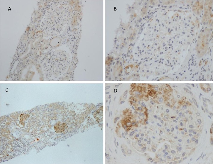
Figure 2.
Renal tissue from patients with IgAN was stained for the presence of C4d. Representative images are shown. (A and B) A patient with a negative C4d staining. (C and D) A representative patient with a positive C4d staining in mesangial areas. No staining is detected in peritubular capillaries. Tubular staining is observed. IgAN, IgA nephropathy. Original magnification, ×100 in A; ×200 in B; ×40 in C; ×400 in D.

