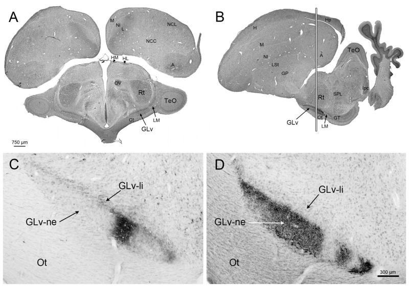
Figure 1. Tectal and retinal terminals in the GLv.
A) Transverse plane of a Giemsa staining showing the position of the GLv in a dorso-ventral orientation. B) Sagittal plane of the Giemsa staining showing the position of the GLv in a rostro-caudal orientation. White vertical line represent aproximatelly the location of the sections in C and D. C) Anterogradely labeled CTB-terminals in the GLv-ne after tectal injection into intermediate layers (injection site not shown). D) Labeled terminals in the Glv-ne after intraocular injection of CTB. Empty areas in the GLv-ne may probably due to uneven distribution of CTB into the vitreal chamber of the eye. Tectal and retinal afferents are topographic and coexist in close apposition into the GLv-ne.

