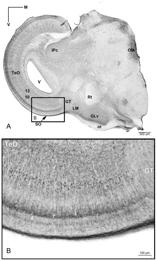
Figure 12. AntiChAT immunohistochemistry in chicken slice.
A) Overview of diencephalic and mesencephalic structures showing antiChAT-DAB labeling. B) Inset of the ventral part of the optic tectum showing anti-ChAT positive cells with the somata located in layer 10 of the TeO. Note that Layer 7 shows neurite immunoreactivity. V = ventricle.

