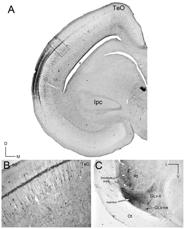
Figure 2. Retrograde tracing of the TeO-GLv projection in vivo.
A) Retrograde CTB-labeling of cells located mainly in layer 10 of the TeO after a GLv injection. B) Higher magnification of the labeled cells located in layer 10 of the TeO. Note the heavy labeled processes in the layer 7. C) Corresponding injection site in the lateral GLv. Orientation in A is the same as B. V = ventral; L = lateral.

