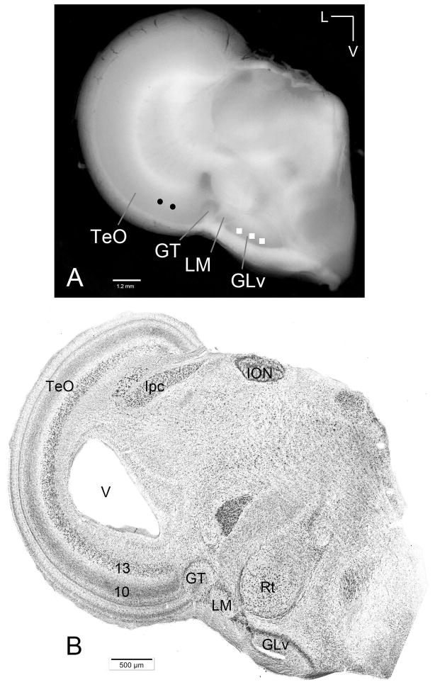
Figure 3. Chicken slice containing the TeO-GLv connection.
A) Macrophotography of a typical 500 μm slice used in this study. Circles and squares mark the location of extracellular injections of dextran amines into the TeO and GLv (performed in each slice) B) Giemsa staining of a 60 μm section showing tectum, pretectum and thalamus.. V = ventricle. Orientation: M = medial; V = ventral.

