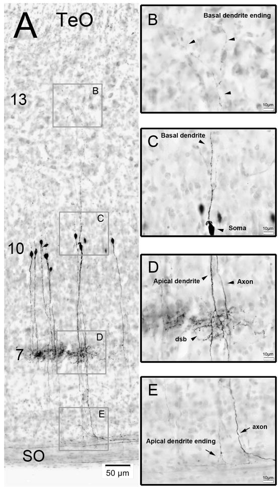
Figure 6. Retrograde labeling of cells in the TeO after BDA injections into GLv.
A) Retrogradely labeled cells with nissl counterstain. Basal dendrites extend until layer 13, somata are located in layer 10, dendritic side-branches lie in layer 7 and apical dendrites extend until layer 2 of the TeO. B) Inset showing the basal dendrite ending in layer 13 (black arrowheads). C) Inset showing the soma and the basal dendrite (black arrowheads). D) Inset showing the apical dendrite, the axon and the dendritic side-branch (dsb) of a neuron (black arrowheads). E) Inset showing the apical dendrite ending and the axon of one vine-neuron. Note the 90° bending of the axons between layer 2 and SO.

