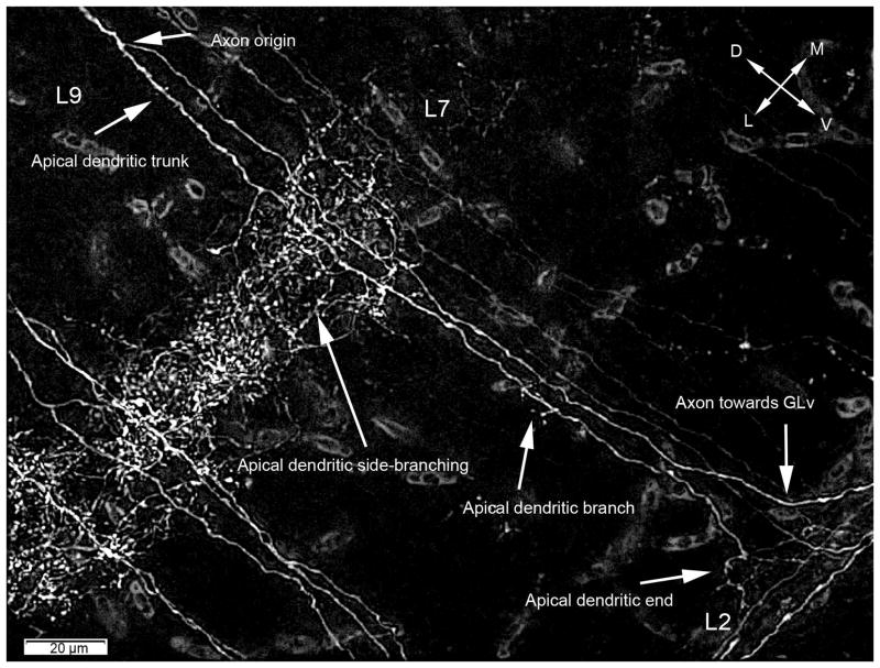
Figure 7. Deconvolution and extended focus projection of a retrogradely labeled cell in the TeO after GLv injection of D-Alexa-546.
Retrograde labeling shows that the axon splits from the apical denritic trunk at the level of layer 9. The apical dendrite side-branch massively in layer 7, continuing with a smaller branching in layer 4. Finally the apical dendrite ends in layer 2. Deconvolution used = Wiener. Orientation: L = lateral; V = ventral.

