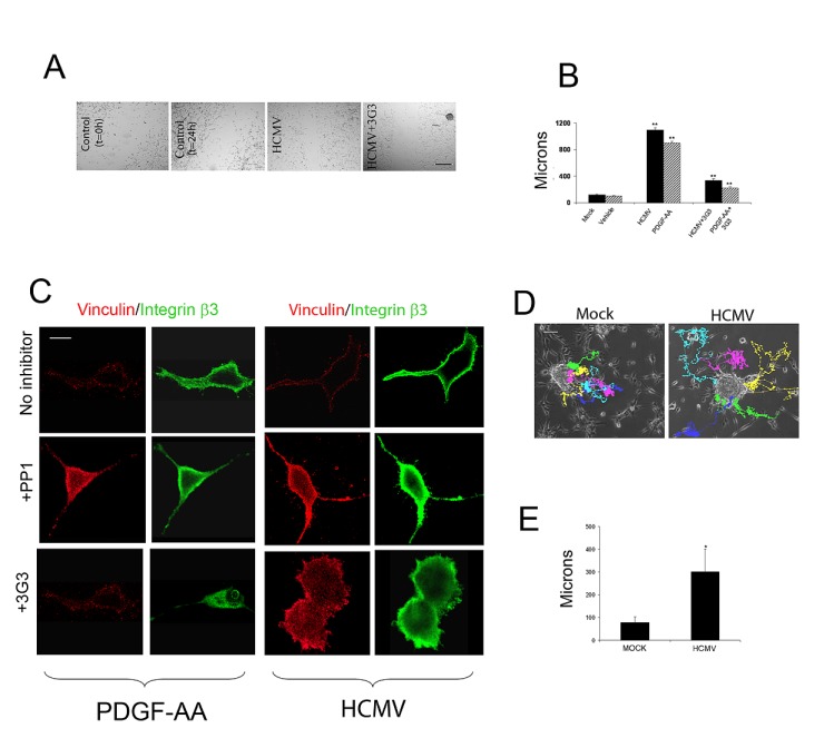
Figure 1. HCMV promotes glioma cell motility and enhances primary glioma stem cell migration.
A. Sixty thousand U87 cells/well were plated onto vitronectin-coated (5 ug/ml) coverslips and cultured overnight. Cells were then serum starved in DMEM and 1% BSA for 24h. The confluent monolayer was scratched with a 1000 ul pipette tip and photographed at 4x magnification under a light microscope immediately after scratching and 24h following the initiation of the “wound closure” process. Cells were pre-treated with blocking agents and stimulated with PDGF-AA, HCMV, or mock, as indicated. B. Quantification of the distance covered by the cells at the edge of the wound. Each condition was run in 6 wells of a 24 well plate and the experiment was repeated twice. *p<0.02, Student t-TEST. C. 30,000 U87 glioma cells were plated onto vitronectin-coated (5 ug/ml) coverslips and serum-starved overnight. Cells were treated with blocking reagents for 30 minutes, stimulated with PDGF-AA (20ng/ml), HCMV (MOI=1), or Mock for 10 minutes and processed for immunofluorescence. Cells were visualized at 60X on a Nikon confocal microscope. PDGF and HCMV treatment induced recruitment of the αvβ3 integrin to focal adhesions in cell cortex (upper rows) and this effect was inhibited by the Src inhibitor PP1 (2.5mM, middle row) and by the PDGFRα blocking antibody 3G3 (10ug/ml, lower panels). D. Tracking of GBM cell migration away from neurospheres growing on surface of cell culture plates under control conditions without HCMV (left panel) or after exposure to HCMV (right panel). Individual cells are indicated by distinct colors. Time indicated in hours. The average total displacement of GBM cells was increased by more than two-fold (unpaired t-test, p<0.001 ).

