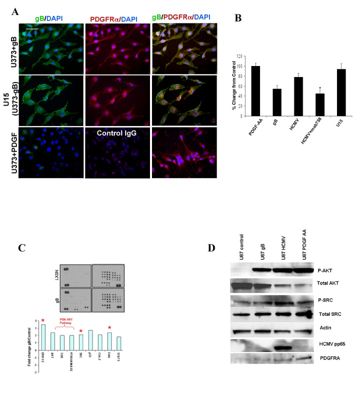
Figure 3. HCMV gB ectopic expression induces activation of the phospho-PDGFRα-PI3K-AKT pathway in human glioblastoma cells.
A.U373 glioma cells grown in serum free conditions (lower panels) stimulated with PDGF-AA (5 ng/ml, 20 min), stably expressing gB (middle panels, U15) or stimulated with recombinant HCMV gB were immunostained for gB (green fluorescence)and phospho-PDGFRα (red fluorescence). Nuclei counterstained with DAPI. gB expressing U15 cells (U373 stably expressing gB) exhibit activation of PDGFRα in the absence of extrinsic stimuli. gB and p-PDGFRα are co-localized at the leading edge of the U15 cells (arrows). Bar= 100μm. B. Quantification of matrigel invasion of U373 glioma and U15 cells stimulated as indicated. Each condition was run in quadruplicate and the experiment was repeated twice. All cells which migrated through Matrigel were stained and counted. A representative experiment is shown. * p=0.01, student t-TEST. C. Glioma cells stably expressing recombinant gB and control (LXSN) were interrogated using a phosphor-kinase proteome profiler array. Densitometry measurements were used to quantify the results, shown in the lower panel. Expression of marked phosphor-proteins were confirmed using Western blot. D. U87 cells were stimulated as shown and processed for western blot using the indicated antibodies.

