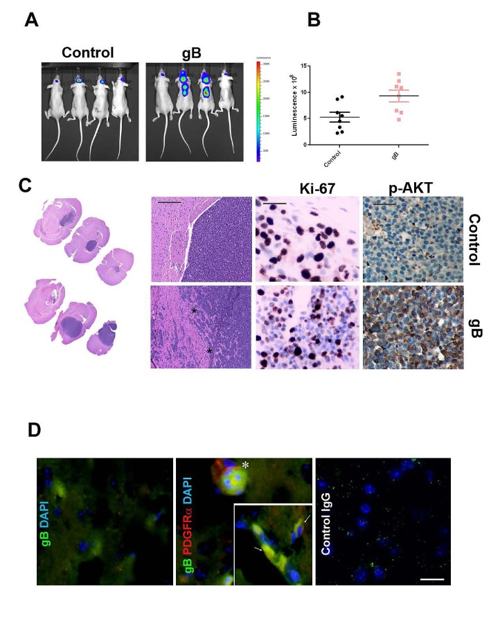
Figure 6. HCMV gB enhances glioblastoma aggressiveness in vivo.
A. Representative bioluminescence images of nude mice bearing U87-Luc intracranial gliomas transduced with gB or control PLXSN vector. Images were acquired 28 days following implantation of 300,000 cells. B. Bioluminescence imaging measurements at day 28 post tumor implantation. C. H&E staining of U87-control (upper left panels) and U87-gB expressing xenograft tissues (lower left panels) at two different levels of magnification (5X and 20X). Bar= 200μm. Middle panels: Ki67 immunostaining of tumor tissue sections. Right panels show IHC detection of p-AKT in consecutive sections from the same tissue samples. Representative images are shown Bar=75μm. D. Double immunofluorescence was used to detect HCMV gB (green fluorescence) and phospho-PDGFRα (red fluorescence) in U87-gB expressing tumor tissue. Inset shows a higher magnification of gB expressing glioma cells which also express p-PDGFRα. Nuclei are stained with DAPI. Bar=100μm.

