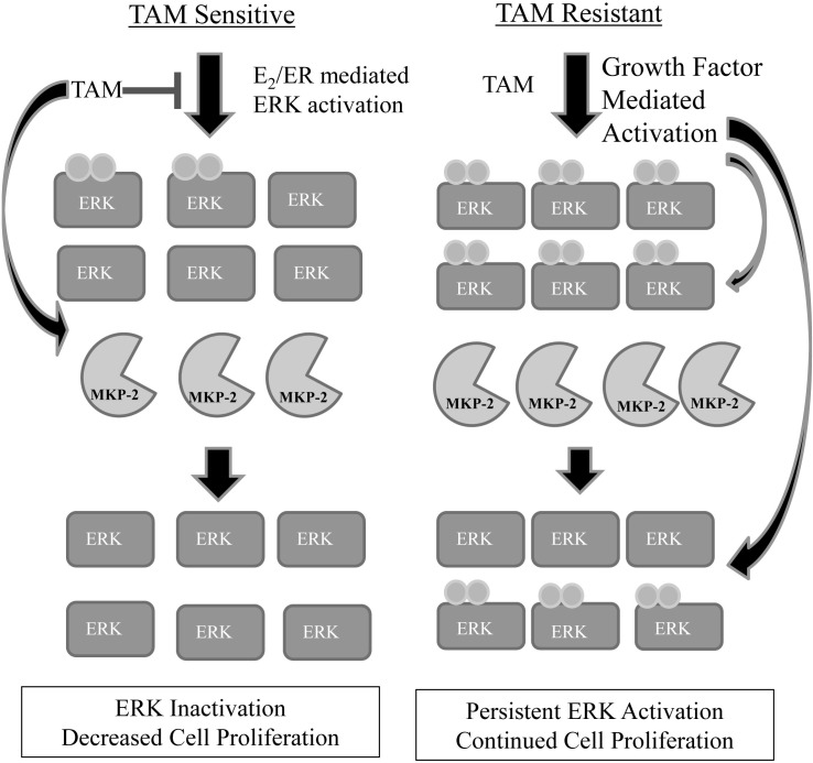
Figure 5. Proposed model of MKP-2 regulation in tamoxifen sensitive vs. tamoxifen resistant cells.
In tamoxifen sensitive cells, low levels of phosphorylated ERK1/2 are present, indicating that cell-growth signaling pathways are activated. Following treatment with TAM, MKP-2 protein expression is increased with resultant dephosphorylation of ERK1/2 and slowing or elimination of cell proliferation. In tamoxifen resistant cells, phosphorylated ERK1/2 is constitutively present at higher levels than in tamoxifen sensitive cells. MKP-2 protein expression is upregulated in an attempt to return phospho-ERK1/2 levels to that of a tamoxifen sensitive cell. The levels of active ERK may be too high for MKP-2 to completely dephosphorylate ERK1/2, resulting in continued cell survival. Additionally, previous work in our lab has shown that active ERK is able to phosphorylate MKP-2, leading to its stabilization, which might be an additional factor in the higher protein levels seen in tamoxifen resistant cells compared to tamoxifen sensitive cells.

