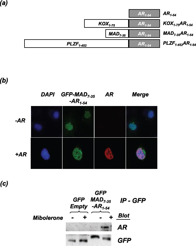
Figure 1. The repressor constructs enter the nucleus and interact with the active androgen receptor.

(a) Schematic representation of the engineered repressors (not drawn to scale). (b) COS-1 cells were transfected with the AR and GFP-MAD7-35-AR1-54. Cells were fixed following 2hrs of treatment with mibolerone. Confocal microscopy was used to visualise the localisation of GFP-MAD7-35-AR1-54 (green) and the full-length AR (stained using ALEXA 594 (red)). Nuclear staining = DAPI (blue). (c) COS-1 cells were transfected with the AR and and GFP-MAD7-35-AR1-54 or GFP-Empty. Cells were treated ± mibolerone for 2hrs and complexes immunoprecipitated with an anti-GFP antibody. Immunoprecipitated complexes were separated using SDS-PAGE and immunoblotted for AR (using an antibody that does not recognise residues 1-54) and GFP.
