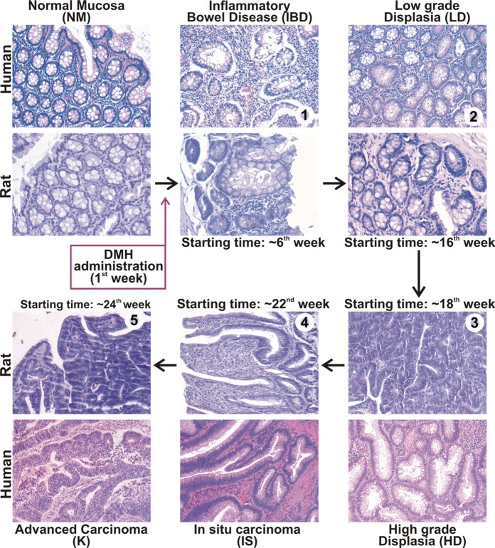
Figure 1. Histology of colonic mucosa in rat model of DMH-induced colorectal cancer.
Resuming scheme reporting the histology of normal mucosa (NM), IBD, dysplasia and colorectal cancer and the timing of the sequential steps during DMH-induced colon carcinogenesis in BDIX rats, compared with human tissues. Sections from rat colon resected from the 6th to the 30th week after the first DMH administration were subjected to histological examination, and compared to human specimens diagnosed as IBD, low grade dysplasia (LD), high grade dysplasia (HD), in situ carcinoma (IS) or advanced carcinoma (K). Colon from untreated animals or normal colon biopsy specimens were used as reference for normal mucosa (NM) morphology.

