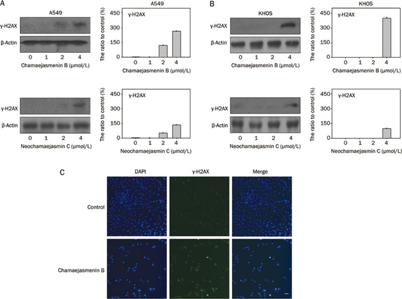
Figure 2.
Chamaejasmenin B and neochamaejasmin C induced DNA damage. (A) A549 cells were exposed to the compounds for 48 h, and protein extracts were immunoblotted for γ-H2AX detection. (B) KHOS cells were exposed to the compounds for 48 h, and protein extracts were immunoblotted for γ-H2AX detection. (C) A549 cells were treated with chamaejasmenin B (2 μmol/L) for 48 h and imaged by immunofluorescence for γ-H2AX (green) and nuclei (blue). γ-H2AX expression is indicated by a punctate appearance in the nucleus. Scale bar=40 μm.

