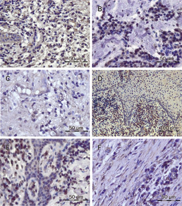
Figure 2.
Photomicrographs of representative iNOS-positive and 3-nitrotyrosine-positive stains in gingival tissue from patients with chronic periodontitis (immunohistochemical staining): (A) countless iNOS+ PMNs and monocytes (scale bar, 50 μm); (B) decreased iNOS+ cells in the SRP+placebo group (scale bar, 50 μm); (C) sparse iNOS+ cells in SRP+SDD group (scale bar, 50 μm); (D) countless 3NT+ PMNs and monocytes (scale bar, 100 μm); (E) decreased 3NT+ cells in the SRP+placebo group (scale bar, 50 μm); (F) sparse 3NT+ cells in the SRP+SDD group (scale bar, 50 μm). This figure is representative of 3 measurements (×400). iNOS+=induced nitric oxide synthase positive cells; 3NT+=3-nitrotyrosine positive cells; SRP=scaling and root planing; SDD=subantimicrobial dose of doxycycline.

