
Figure 1. Killing assay.
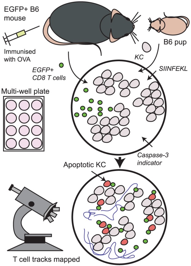
Primary KC from B6 mouse pups were cultured in multi-well plates until 70% confluent, and then loaded with SIINFEKL peptide. Antigen-specific CD8+ T cells from EGFP+ B6 mice were isolated and co-cultured with target cells in the presence of cell-permeable caspase-3 indicator dye and imaged by time-lapse fluorescence microscopy or by confocal microscopy every 12 minutes for 30 h. Cell death was noted by colour change, morphology, and behaviour.