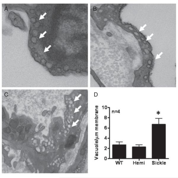
Figure 3.
Ultrastructure of the endothelium using transmission electron microscopy in wild type (WT) (A), heterozygous (Hemi; B), and transgenic sickle-cell (Sickle; C) mice penes. Magnification 40,000×. Arrows denote vacuoles. (D) Quantitative assessment of vacuole density per μg membrane length. Measurements were made in blinded fashion. Images are representative of four penile samples. n indicates number of experiments. *P < 0.05 vs. WT and Hemi.

