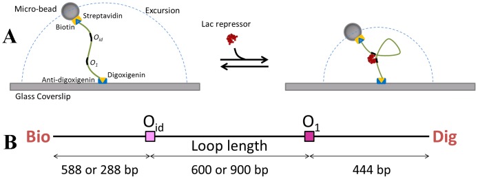
Figure 7. Illustrations of the TPM experiment and the 1632 bp DNA.

A. DNA tether labeled with biotin and digoxigenin links a polystreptavidin-coated microsphere (bead) to an anti-digoxigenin coated coverslip. The motion of this tethered bead is characterized by its mean (or RMS) excursion, which exhibits a visible decrease when subject to the formation of Lac repressor mediated loop. B. Schematic linear representation of the 1632 bp DNA construct with Oid and O1 positioned 600 or 900 base pairs apart.
