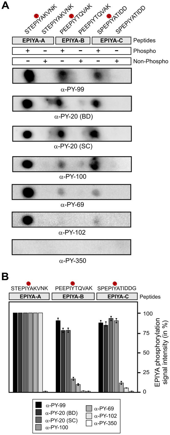
Figure 2. Variable recognition of synthetic 11-mer CagA phospho-peptides by seven commercial α-phosphotyrosine antibodies.

(A) Dotblot analysis of the indicated phospho- and non-phospho peptides derived from single EPIYA-motifs A, B and C. All Dotblots were probed with the indicated commercial phosphotyrosine antibodies and exposed as described in the Material & Methods section. (B) Quantification of spot intensities on Dotblots. Signal intensities were measured densitometrically with the Lumi-Imager F1 and revealed the percentage of phosphorylation signal per sample. The strongest spot on every Dotblot was set at 100% for each of the different α-phosphotyrosine antibodies as indicated. Quantitation results are shown for three independent experiments.
