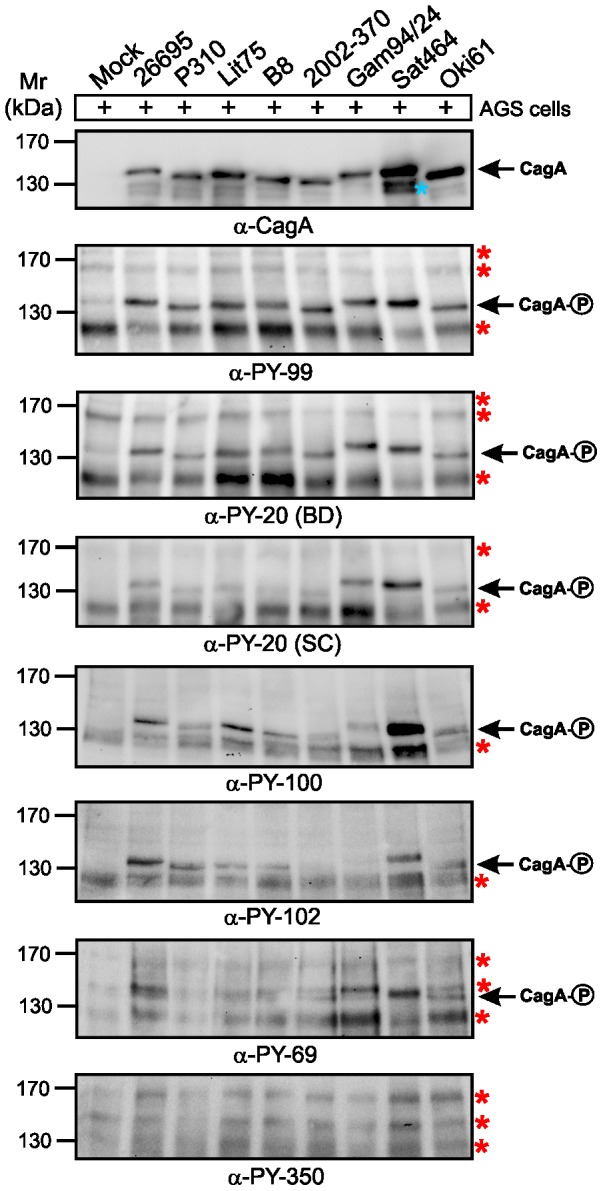
Figure 5. Role of EPIYA motifs in CagA phosphorylation during H. pylori infection was investigated with seven different α-phosphotyrosine antibodies.

AGS cells were infected for 6-expressing H. pylori strains as indicated. The samples in Figure 4 were harvested after photographing. Phosphorylation of CagA was examined using the indicated α–phosphotyrosine antibodies. Loading of equal amounts of CagA from each sample was confirmed by probing with a monoclonal α-CagA antibody. A larger section of the ∼120−180 kDa range is shown and contains the phospho-CagA bands of different sizes (arrows) as well as a set of tyrosine-phosphorylated host cell proteins (red asterisks). The blue asterisk indicates a putative N-terminal fragment of CagA which sometimes appears on SDS-PAGE gels [23].
