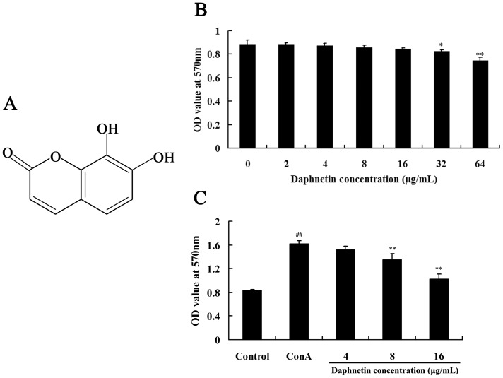
Figure 1. Effect of daphnetin on mouse splenocytes viability and proliferation.
(A) Chemical structure of daphnetin used in the study. (B) Effect of daphnetin on the viability of mouse splenocytes. The cells were treated with daphnetin (0–64 µg/mL) for 48 h. The cell viability was determined by MTT assay. (C) Effect of daphnetin on the ConA induced mouse splenocytes proliferation. Splenocytes cultured with fisetin (4, 8, 16 µg/mL) combined with ConA (5 µg/mL) for 24 h. Cell proliferation was assessed by MTT assay. Data are presented as means ± SD of three independent experiments. Significant differences from control group were indicated by *P<0.05 and **P<0.01.

