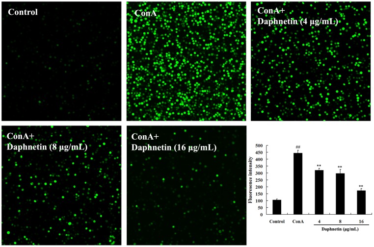
Figure 4. Effects of different concentration of daphnetin on the [Ca2+]i in mouse T cells.
T Cells were pretreated with 20 µM Fluo-3-AM and incubated in the presence of daphnetin (4, 8, 16 µg/mL) for 30 min at 37°C and measured at 37°C using a Confocal Laser Microscope. The fluorescence intensity in ConA group was increased compared with the control group, and daphnetin could diminish the fluorescence intensity. The results were from three independent experiments and presented as mean ± SD. ## P<0.01 vs. Control group; **P<0.01 vs. ConA group.

