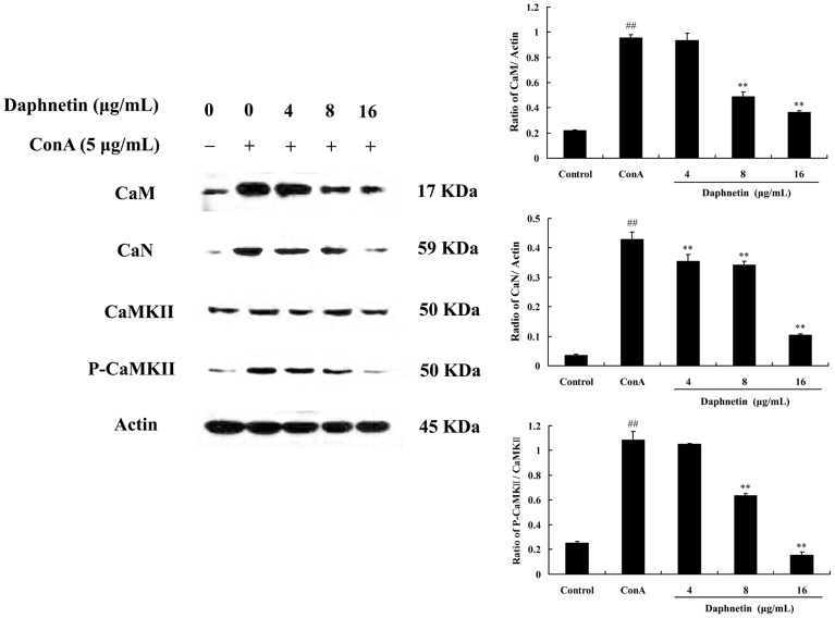
Figure 5. Effect of daphnetin on [Ca2+]i -related signaling pathway in mouse T cells.
Mouse CD3 T cells were treated with daphnetin (4, 8, 16 µg/mL) for 0.5 h and then stimulated with ConA (5 µg/mL) for 0.5 h. Representative western blots showed protein expression of CaM, CaN, CaMKII and P-CaMKII. Data were presented as means ± SD of three independent experiments. ## P<0.01 vs. Control group; **P<0.01 vs. ConA group.

