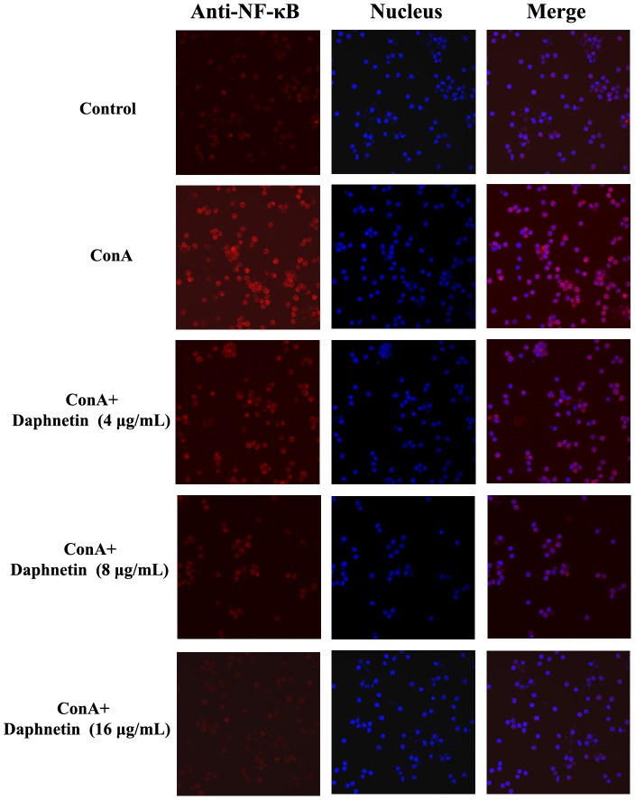
Figure 7. Influence of daphnetin on NF-κB translocation in mouse T cells.
Mouse CD3 T cells were treated with 4, 8 and 16 µg/mL of daphnetin for 1 h and then stimulated with ConA for 30 min. Control group: NF-κB was localized to the cytoplasm; ConA group: Cells treated with ConA showed nuclear distribution of NF-κB; Daphnetin group: The localization of NF-κB was mainly cytoplasmic, demonstrating a reduction in the ability to translocate NF-κB. The fluorescence images were recorded with an Olympus Confocal Laser Scanning Biological Microscope.

