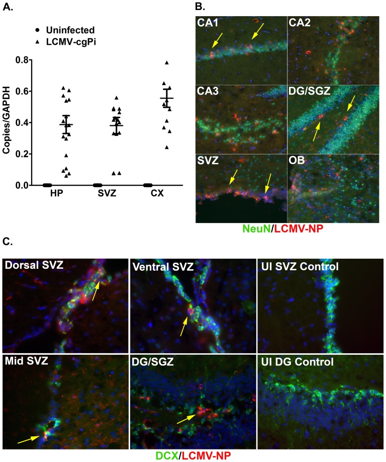
Figure 1. LCMV infects cells in the hippocampus and subventricular zone.
In 6-week old LCMV-cgPi mice, (A) RT-QPCR quantification of LCMV-glycoprotein RNA in hippocampus (HP), subventricular zone (SVZ), and cortex (CX) is shown with mean ± SEM; N≥10 mice. Uninfected controls were included in the experiment to demonstrate primer specificity. (B) Representative IHC images show infection of neurons (NeuN+ cells) in the SVZ, olfactory bulb (OB), and regions of hippocampus: subgranular zone (SGZ) of dentate gyrus (DG) and CA1, CA2, and CA3. Tissue sections were stained with anti-LCMV-nucleoprotein and anti-NeuN. Table 1 shows quantitative data for levels of infection. (C) Representative IHC images show infection of neuroblasts (DCX+ cells) in the SVZ and SGZ. Tissue sections were stained with anti-LCMV-nucleoprotein (NP) and anti-DCX. Uninfected controls were included for all IHC experiments that used the anti-LCMV-nucleoprotein antibody.

